Current Studies
SENSory Evaluation and Instrument Testing (SENSE-IT): An evaluation of psychometric quality of an electrical perceptual sensory device
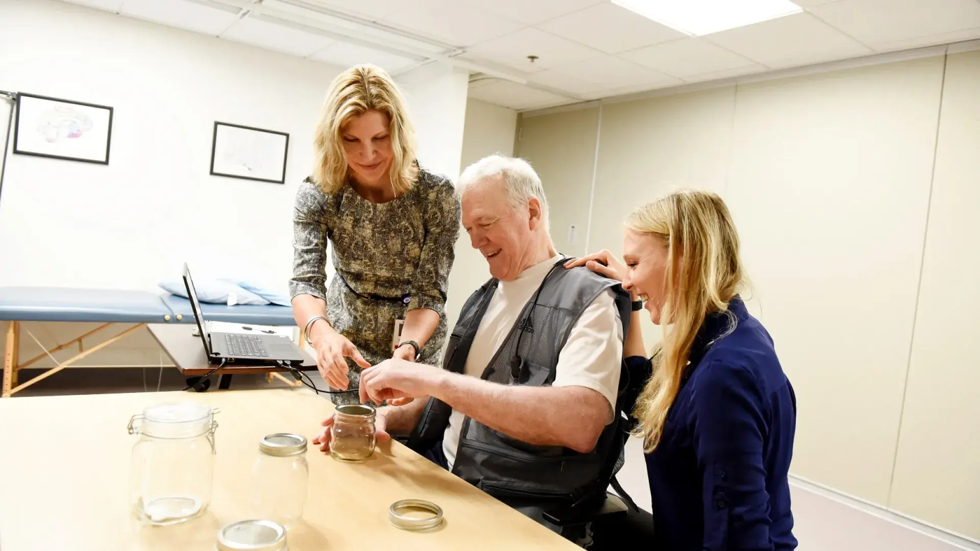
Sensory impairments are common across various clinical disorders, including, but not limited to, diabetes, stroke, spinal cord injury, multiple sclerosis, peripheral nerve injuries, and amputation. Additionally, sensory loss is a natural consequence of aging. However, a lack of reliable, valid, and sensitive somatosensory evaluation tools limits the ability to precisely measure impairment or changes in sensory perception.
We are seeking healthy adults and those with impaired sensation to participate in this research study investigating the reliability and validity of electrical stimulation as a method of measuring sensation. Electrical stimulation is a safe and painless tool frequently use din physical therapy practice for a variety of purposes.
DYT1 genotype- and phenotype-specific brain circuits in dystonia
TOR1A-associated (DYT1) dystonia is the most prevalent genetic form of a disorder called primary torsion dystonia and is the third most common movement disorder after Parkinson’s disease and essential tremor. Dystonia is a debilitating brain disorder that can affect any body part leading to involuntary, abnormal posturing and pain which can impair nearly all activities of daily living. A variation in one copy of the TOR1A gene leads to symptoms of dystonia in ~30% of people who carry the gene, while the remaining ~70% do not develop dystonic symptoms. The purpose of this study is to learn more about the brain network activity associated with the TOR1A gene.
In collaboration with the Dystonia Partners Research Bank [link], we are recruiting participants with 1) DYT1 dystonia; 2) families who are carriers of DYT1 dystonia, including at least one non-manifesting and one dystonia-manifesting individual; and 3) healthy controls. This study involves an MRI scan, a neurologic exam visit, and a blood sample. Being able to distinguish between people who will develop the disease from those who won't develop the disease will help us to understand the factors that contribute to the development of dystonia. The long-term goal is to understand the disordered process that causes dystonia and to develop sensitive testing and new ways to treat this condition.
The effects of neural modulation on phonatory function in laryngeal dystonia
Laryngeal dystonia (LD) is a disorder that causes uncontrolled spasms of the laryngeal muscles during speech, causing severe speaking difficulty and a reduction in quality of life. Although the cause of the disorder is not fully understood and there are no known treatments for LD that produce long-term benefits, recent findings have discovered that laryngeal dystonia is associated with a lack of neural inhibition that may be linked to the spasms. The purpose of this study is to determine if repetitive transcranial magnetic stimulation (rTMS) is effective for increasing neural inhibition, enhancing voice quality and function, and improving quality of life for people with laryngeal dystonia. rTMS is a safe, noninvasive, and painless procedure that has been shown to regulate excitability by increasing inhibition in the brain. It is our hope that the outcomes of this study will facilitate the development of appropriate stimulation parameters to treat laryngeal dystonia, including identification of who may respond to this type of treatment. We are recruiting participants with LD for this study.
This double-blinded, randomized study occurs in two phases: (1) a 5-day phase of rTMS intervention delivered to the left-brain area, and (2) a 5-day control phase where research staff deliver a sham treatment to the same brain area. There is an approximate 3 month waiting period in between phases.
Stroke Rehabilitation
Impaired use of the arm and hand are a major source of disability for people after a stroke. In the Brain Recovery Lab, we are examining novel ways to enhance recovery after stroke. We recently completed an investigation of the effectiveness of Vagus nerve stimulation (VNS) paired with intensive arm rehabilitation. Results from this multi-site, triple blinded, sham controlled pivotal trial (https://www.clinicaltrials.gov; NCT03131960) found greater clinically meaningful arm improvement for participants receiving VNS paired with intensive therapy compared to participants receiving the control condition. VNS therapy has now been FDA-approved and is being clinically implemented at Massachusetts General Hospital in partnership with Spaulding Rehabilitation Network. The treatment involves a simple outpatient surgical procedure to implant a small VNS stimulator just beneath the chest skin. Following implantation, individuals will receive 18 sessions of intensive arm training over six weeks. The procedure and therapy are covered by Medicare and most insurance plans.
If you are interested in VNS therapy or would like to learn more, please contact the MGH Neurorecovery Clinic at (617) 726-8459.
A multimodal assessment of neurophysiology in focal dystonia
Focal dystonia is a neurologically based movement disorder that can affect any body part, severely impairing a person’s ability to function in their daily life. People with focal dystonia experience involuntary muscle contractions in body areas such as the larynx, hand, neck, or other muscles. Focal dystonias are the third most common movement disorder, yet the disease has no diagnostic test and patients are often misdiagnosed and receive years of unnecessary treatment. We are recruiting participants with focal hand or laryngeal dystonia as well as healthy controls.
The study investigates connections between brain areas using two different types of measures to understand the difference between people with focal dystonia and healthy people, transcranial magnetic brain stimulation (TMS) and brain imaging techniques. TMS causes magnetic fields in the brain to form electrical currents. The weak stimulation allows researchers to measure the excitability of portions of the brain. A brain navigation system will use MRI scans of the brain like a map to target specific areas in the brain. The purpose of the study is to use MRI to determine the structure of the brain while also using TMS to measure your brain activity. Using this information, differences between healthy subjects and those with focal dystonia may be identified and lead to a better understanding of the disorder.
Recruitment for this study has ended.